List of members |
Facilities |
Internships and jobs |
PhD |
Publications |
News |
Team
- Permanent members: Tristan Cren, Pascal David, François Debontridder, Marie Hervé
Since its foundation in the early 80’s, our team has been designing, developing and perfecting original experimental sets at the cutting edge of nano-technologies. Thus, the first French scanning tunneling microscope (STM) operating at low temperature (M1) was born in the laboratory in 1993, followed by the first “all-digital” low temperature STM (M2) in 2000. The exceptional performance in tunneling spectroscopy of this second microscope paved the way in 2004 for Josephson microscopy, using superconducting tips. Our third microscope (M3), the result of 9 years of development, was inaugurated in 2008. This device combines a very low temperature STM (300mK) operating under ultra-high vacuum and strong magnetic field with a chamber for in-situ sample preparation and characterization.
Our current developments include a new low temperature cryogen free STM/AFM and a radio frequency STM that will be implemented on M3. In order to strengthening the capacities of our instrumental park, we are also pursuing the development of several ultra-high vacuum systems or sub-assemblies: 4-axis cryogenic manipulators, evaporation chamber, etc… We have also commissioned a preparation chamber coupled with a variable temperature STM in order to increase our sample preparation capacities.
- Radio frequency Scanning Tunneling Microscope
- 2K AFM/STM UHV with 2T/1T vector magnetic field
- UHV suitcase with organic molecules evaporation facility
Radio frequency Scanning Tunneling Microscope
In condensed matter systems, the investigation of low energy magnetic excitations has been carried out using resonance method such as Electron Paramagnetic Resonance (EPR), Nuclear Magnetic resonance (NMR) or Ferromagnetic Resonance (FMR) [1]. Conventional magnetic resonance techniques are based on the inductive detection of an electromagnetic signal induced by the precession of an electron’s or nucleus’s magnetic moment and require investigating macroscopic systems. With the development of low dimension electronic and quantum computing, one challenge in condensed matter physics is to develop radio frequency (rf)-based resonance experiments liable to sense low energy electronic, magnetic and vibronic excitations down to the single atom / molecule limit.
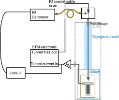
Scanning tunnelling microscopy (STM) is an ideal tool for investigating electronic, magnetic, as well as vibronic excitations of single molecules or atoms on [2-4] surfaces in the sub-meV range. Indeed, such excitation spectrum can be probed using conventional STM as long as the energy separation between the states is larger than 3.2kbT, i.e., the Fermi-Dirac broadening at finite temperature. To give an example, at a temperature of 300 mK, the STM energy resolution is about 90 µeV. It can be enhanced down to 30 µeV with the use of a superconducting tip. To go further, a possibility would be to use a resonance technique: if combined to STM, it is liable to address electronic, magnetic and vibronic excitation spectrum down to few nano-eVs resolution at the atomic scale. Access to such an extreme energy resolution and high frequency signal in STM was a long-standing problem. Conventional STM has a natural limitation to detect high frequency signal. This is due to the bandwidth of its transimpedence amplifier used to convert tunnelling current in the pA / nA range to manageable voltage. Typical bandwidths are around several kHz which is far too low to track high frequency signal corresponding to µeV energy splitting.
Since a few years a new promising method allows to go much further by exploiting radio-frequency resonances [5-7]. The recently developed rf-STM, it is liable to address electronic, magnetic and vibronic excitation spectrum down to few nano-eVs resolution at the atomic scale. The possibility to access such a high energy resolution with an STM was demonstrated only recently in the literature and such an experimental setup is currently under construction at INSP.
[1] C. P. Slichter Principles of Magnetic Resonance 1996). [2] M. F. Crommie, C. P. Lutz, and D. M. Eigler, Science 262, 218 (1993). [3] A. J. Heinrich, J. A. Gupta, C. P. Lutz, and D. M. Eigler, Science 306, 466 (2004). [4] J. K. Gimzewski, E. Stoll, and R. R. Schlittler, Surface Science 181, 267 (1987). [5] M. Herve, M. Peter, and W. Wulfhekel, Applied Physics Letters 107, 093101 (2015). [6] S. Baumann, W. Paul, T. Choi, C. P. Lutz, A. Ardavan, and A. J. Heinrich, Science 350, 417 (2015). [7] M. Hervé, M. Peter, T. Balashov, and W. Wulfhekel, Nanomaterials 9, 827 (2019).Main collaborations
- Wulf Wulfhekel (Karlsruhe Institute of Technology)
Founding
- Projet ANR Ginet2.0
- Projet Emergence MAGRES
- Credits CNRS DIALOG
- Appel à projet spécifique de l’INP
Publications récentes
- M. Hervé, M. Peter, T. Balashov, and W. Wulfhekel.Towards Laterally Resolved Ferromagnetic Resonance with Spin-Polarized Scanning Tunneling Microscopy. Nanomaterials 9, 827 (2019) https://doi.org/10.3390/nano9060827
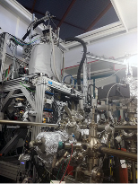
2K AFM/STM UHV with 2T/1T vector magnetic field
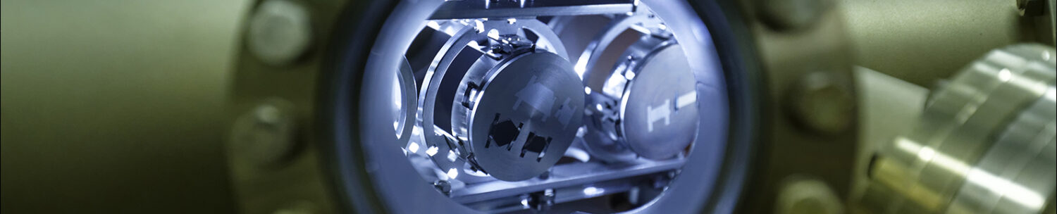
A new generation UHV AFM/STM operating at 2K in a 2T vertical 1T horizontal magnetic field is currently under development. This equipment is derived from a UHV system “Omicron LT” whose microscope and cryogenic system are developed in the laboratory. The originality of this new set-up lies in the use of a “cryogen free” cryogenic system with a pulsed tube refrigerator coupled to a 1K pot. This cryogenic system avoids the use of liquid helium and the interruptions linked to the filling of the cryostat.
 To prevent mechanical noise produced by the coupling of the pulsed tube of the cryogenerator with the STM, an anti-vibration system with a double suspension mechanism has been developed. Fully interfaced, the instrument is remotely controllable and will allow to record highly resolved tunnel spectroscopy maps by varying the temperature and/or the magnetic field over very long times. The microscope head is equipped with 6 independent contacts on the sample allowing operando measurements on devices, as well as 6 independent contacts on the tip of the microscope, allowing to operate in STM or NC-AFM mode, or to perform resistivity measurements on thin films elaborated in-situ.
© INSP
UHV suitcase with organic molecules evaporation facility
The team has developed a modular chamber allowing the evaporation of organic molecules under ultra-high vacuum conditions (P<10-10mbar) on samples elaborated in the preparation chambers of our STM set-ups. This way, we avoid polluting our chambers with organic molecules while keeping all the growth process and analysis of samples under UHV.
This mini preparation chamber is equipped with a Joule heating stage and a cold finger. It gives access to 100-800K temperature range. Viewports allows to follow-up by optical spectroscopy in the UV-visible range the growth of evaporated layers. Its small size and its autonomous pumping by NEG make it a vacuum suitcase allowing the samples to be transported on different systems under UHV.
© INSP
We have extensively used it for the study of local properties of magnetic states induced on the surface of superconductors by one- and two-dimensional arrays of molecular magnets in the framework of Danilo Longo’s PhD thesis. Since its commissioning in 2017, this chamber has been operated on 3 set-ups in the laboratory as well as on the DEIMOS beamline at synchrotron SOLEIL.
© INSP