List of members |
Facilities |
Internships and jobs |
PhD |
Publications |
News |
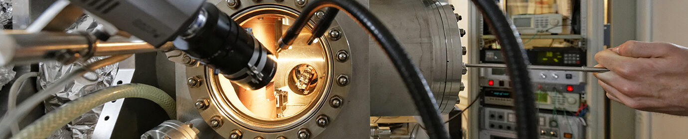
Reflection polarization microscope for imaging magnetic domains by magneto-optical Kerr effect
- Contact-cooled sample in an Oxford Microstat BT HiRes II pillar cryostat (4-300 K)
- Various light sources: filtered white lamp, LED, continuous laser (Ti-sapphire, Argon, He-Ne)
- Andor Clara CCD camera detector with brightness intensifier, with the possibility of external triggering of the image acquisition.
It is also possible to perform micro-spectroscopy experiments by selecting the signal of a part of the image and analyzing it in a spectrometer.
Magnetic fields perpendicular to the plane :
- electromagnet delivering up to 60mT
- superconducting coil delivering up to 6T
- in-house micro-coil installed on the sample, in the cryostat, delivering up to 200mT, rise time 140ns
Planar magnetic fields
- air-cooled CAYLAR rotating dipole delivering up to 500mT (Fig. 2b)
- in-house solenoid delivering up to 50mT
- home-made micro-coil mounted in the cryostat, on the cold finger, delivering up to 15 mT, rise time 150ns (Fig. 2a)
Polar Kerr effect configuration
- Detection of the magnetization component perpendicular to the sample
- Domain and domain wall studies of out-of-plane magnetized layers: Dourlat et al., PRB ’08, Gourdon et al., PRB ’07, Gourdon et al., PRB ’09, Thevenard et al., PRB ’11, Haghgoo et al., PRB’10
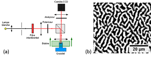
Figure 1 :(a) Scheme of the Kerr polar experimental device (scheme A. Dourlat). (b) Self-organization in zero field of domain walls in (Ga,Mn)(As,P) magnetized out of plane Haghgoo et al., PRB’10.
Longitudinal Kerr effect configuration
- Detection of the magnetization component in the sample plane
- Study of domains and domain walls of magnetized layers in the plane: Thevenard et al.PRB ’12, Tortarolo et al., APL’12
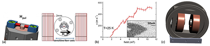
Caption 2 : (a) Micro-coil to be inserted in the cryostat to obtain short magnetic field pulses in the sample plane. The external coils (red) allow to go to higher fields. (b) Propagation of domain walls measured on GaMnAs magnetized in the plane. Thevenard et al. PRB ’12 (c) CAYLAR rotating dipole (33mm air gap).
Contacts
- Catherine Gourdon : catherine.gourdon(at)insp.jussieu.fr
- Laura Thevenard : Laura.Thevenard(at)insp.jussieu.fr